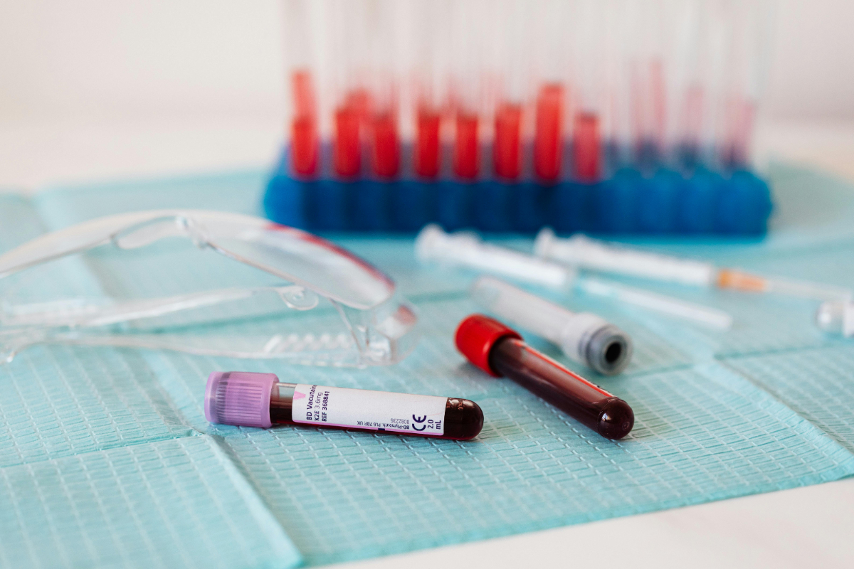
Most people who receive a complete blood count report focus on the numbers they recognize — hemoglobin, white blood cells, platelets. Then their eyes land on a cluster of unfamiliar abbreviations: MCV, MCH, MCHC, and RDW. One or more is flagged outside the reference range. A quick internet search returns dense medical jargon about "microcytic hypochromic anemia" and "anisocytosis," and suddenly a routine lab report feels impenetrable.
Here is the simple truth: these four values describe the size, hemoglobin content, and uniformity of your red blood cells. Together, they answer a question that hemoglobin and hematocrit alone cannot: if anemia is present — or developing — what is causing it? Knowing the cause determines the treatment. Taking iron pills when you actually need vitamin B12 is not just ineffective — it can be harmful. Understanding red cell indices protects you from that mistake.
Disclaimer: This article is for educational purposes only and does not replace professional medical advice. Always consult a qualified healthcare provider for diagnosis and treatment.
What Are Red Cell Indices?
Think of your bloodstream as a fleet of delivery trucks. Red blood cells (RBCs) are the trucks, and hemoglobin is the cargo — oxygen bound to iron. Knowing the total number of trucks (RBC count) and the total cargo delivered (hemoglobin) gives you a broad picture. But red cell indices tell you about the trucks themselves:
- MCV — How big is each truck?
- MCH — How much cargo does each truck carry?
- MCHC — How tightly is the cargo packed in each truck?
- RDW — Are all the trucks the same size, or is the fleet a chaotic mix of small and large vehicles?
These parameters are calculated automatically by hematology analyzers at every major lab (Quest Diagnostics, LabCorp, Synlab) and cost nothing extra — they are part of every standard CBC.
MCV — Mean Corpuscular Volume
What it measures: The average volume (size) of a single red blood cell, expressed in femtoliters (fL).
Normal range (CLSI): 80–100 fL
MCV is the single most useful red cell index for classifying anemia. It divides anemias into three categories:
Low MCV (below 80 fL) — Microcytic Anemia
Red blood cells are abnormally small. In clinical practice, the overwhelming majority of low-MCV cases are caused by:
-
Iron deficiency anemia — the most common anemia worldwide. When iron stores are depleted, the bone marrow cannot produce enough hemoglobin to fill each red cell. It compensates by making smaller cells. This is particularly common in menstruating women, pregnant women, growing adolescents, and individuals with chronic blood loss (GI bleeding, heavy periods). Check iron and ferritin levels for confirmation.
-
Thalassemia trait — an inherited condition common in people of Mediterranean, Southeast Asian, Middle Eastern, and African descent. The bone marrow produces structurally abnormal hemoglobin chains, resulting in small red cells. Unlike iron deficiency, ferritin is normal or elevated. A hemoglobin electrophoresis test distinguishes the two.
-
Anemia of chronic disease — long-standing inflammatory conditions (rheumatoid arthritis, Crohn's disease, chronic kidney disease) can produce a mildly microcytic picture, though normocytic anemia (normal MCV) is more typical.
-
Lead poisoning — rare in adults but seen in occupational exposure and in children exposed to lead paint.
Normal MCV (80–100 fL) — Normocytic Anemia
Red blood cells are normal in size, but there are too few of them or they carry too little hemoglobin. Causes include:
- Acute blood loss (surgery, trauma, GI bleed).
- Anemia of chronic disease (early or moderate stage).
- Chronic kidney disease (reduced erythropoietin production).
- Hemolytic anemias (red cells destroyed prematurely).
- Mixed deficiencies (simultaneous iron and B12 deficiency can average out to a normal MCV).
High MCV (above 100 fL) — Macrocytic Anemia
Red blood cells are abnormally large. The two most important causes:
-
Vitamin B12 deficiency — B12 is essential for normal DNA synthesis during red cell maturation. Without it, cells cannot divide properly in the bone marrow; they grow larger and larger without splitting, producing oversized red cells (megaloblasts). Symptoms include fatigue, numbness or tingling in the hands and feet, memory problems, and mood changes. At-risk groups: vegans, vegetarians, adults over 60 (reduced stomach acid impairs B12 absorption), and patients on metformin or proton pump inhibitors. See our vitamin blood test guide for more detail.
-
Folate (B9) deficiency — produces an identical blood picture to B12 deficiency. Common in pregnancy (increased demand), alcoholism, and diets extremely low in leafy greens.
-
Alcohol use — even without overt deficiency, chronic alcohol consumption directly impairs red cell maturation, raising MCV. In fact, an elevated MCV in an otherwise healthy-appearing adult is sometimes the first laboratory clue to heavy alcohol intake. For more on this topic, see nicotine and alcohol effects on blood tests.
-
Hypothyroidism — low thyroid function slows bone marrow metabolism, producing mildly macrocytic cells. If MCV is elevated alongside fatigue and weight gain, check thyroid tests.
-
Medications — methotrexate, hydroxyurea, azathioprine, and some antiretroviral drugs interfere with DNA synthesis and raise MCV.
-
Myelodysplastic syndromes — rare bone marrow disorders in older adults that produce large, abnormal red cells.
MCH — Mean Corpuscular Hemoglobin
What it measures: The average amount of hemoglobin in a single red blood cell, expressed in picograms (pg).
Normal range: 27–33 pg
MCH moves in near-lockstep with MCV:
- Low MCH (below 27 pg) — the cell is small and carries less hemoglobin than normal. This is the hallmark of iron deficiency. The red cell is literally underfilled — like a delivery truck running half-empty because there was not enough cargo at the warehouse.
- High MCH (above 33 pg) — the cell is large and overstuffed with hemoglobin. This accompanies B12 or folate deficiency. However, the oversized cell is fragile and gets destroyed faster in the spleen, which is why patients still feel anemic despite apparently "hemoglobin-rich" cells.
Clinical tip: If MCH is low but hemoglobin is still in the normal range, you may have early iron depletion that has not yet progressed to frank anemia. This is a valuable early warning — especially for women experiencing chronic fatigue or hair loss.
MCHC — Mean Corpuscular Hemoglobin Concentration
What it measures: How densely hemoglobin is packed within each red blood cell, expressed in g/L or g/dL.
Normal range: 320–360 g/L (32–36 g/dL)
MCHC is the most stable of the four indices and rarely deviates from normal in mild conditions.
- Low MCHC (below 320 g/L) — indicates advanced iron deficiency. By the time MCHC drops, the iron depletion is usually severe and long-standing. The cells are not just small (low MCV) and underfilled (low MCH) — they are also dilute, meaning hemoglobin concentration within the cell itself is reduced.
- High MCHC (above 360 g/L) — genuinely elevated MCHC is rare and has a narrow differential:
- Hereditary spherocytosis — a genetic condition where red blood cells lose their normal disc shape and become spheres. The reduced surface-to-volume ratio concentrates hemoglobin.
- Cold agglutinins or lipemic samples — technical artifacts that falsely elevate MCHC. If the value is unexpectedly high, the lab may need to repeat the test or warm the sample.
Because MCHC is so stable, an abnormal result should always be taken seriously and verified with a repeat test.
RDW — Red Cell Distribution Width
What it measures: The degree of variation in red blood cell sizes (anisocytosis), expressed as a percentage (coefficient of variation of the RBC volume distribution).
Normal range: 11.5–14.5%
RDW is arguably the most clinically underrated of the four indices. It answers a simple but powerful question: are your red cells uniform, or are they a mix of sizes?
Why RDW Matters: The Early Warning System
In healthy blood, nearly all red cells are roughly the same size, and RDW stays below 14.5%. When the bone marrow begins struggling — running low on iron, B12, or folate — it starts producing cells of inconsistent size. Some come out too small, some too large, and RDW creeps upward.
This happens before hemoglobin drops. An elevated RDW with a still-normal hemoglobin is one of the earliest detectable signs of developing anemia. It is also valuable for distinguishing between similar-looking conditions:
- Iron deficiency anemia: MCV low + RDW high (mixed cell sizes because old normal-sized cells coexist with new iron-starved small cells).
- Thalassemia trait: MCV low + RDW normal (the bone marrow consistently produces uniformly small cells because the defect is genetic, not nutritional).
This single distinction — RDW high vs normal — can save patients from unnecessary and expensive hemoglobin electrophoresis testing in many cases.
RDW in Other Conditions
- Mixed deficiency (iron + B12 simultaneously): MCV may be normal (small and large cells average out), but RDW will be markedly elevated, revealing the hidden problem.
- Post-transfusion: Receiving donor blood introduces cells of a different size, transiently raising RDW.
- Reticulocytosis: When the bone marrow is actively recovering (after blood loss or treatment of anemia), young red cells (reticulocytes) are larger than mature ones. See reticulocyte count guide for more detail.
- Recent research has linked chronically elevated RDW to cardiovascular disease risk and all-cause mortality, independent of anemia — though the clinical use of this finding is still evolving.
Putting It All Together: Anemia Classification by Indices
The power of red cell indices lies in their combined pattern. Here is a practical decision framework:
| Pattern |
Most Likely Cause |
Next Step |
| MCV low + MCH low + MCHC low + RDW high |
Iron deficiency anemia |
Check ferritin, serum iron, TIBC |
| MCV low + MCH low + MCHC normal + RDW normal |
Thalassemia trait |
Hemoglobin electrophoresis |
| MCV high + MCH high + RDW high |
B12 or folate deficiency |
Check B12, folate, homocysteine |
| MCV high + MCH high + RDW normal |
Liver disease or hypothyroidism |
Liver function, thyroid panel |
| MCV normal + RDW high |
Mixed deficiency or early iron depletion |
Ferritin, B12, folate |
| MCV normal + RDW normal + hemoglobin low |
Acute blood loss or chronic disease |
Clinical context, reticulocyte count |
| All indices normal |
No red cell abnormality |
No further workup needed for anemia |
This table is a guide, not a replacement for clinical judgment. Overlapping conditions (e.g., iron deficiency plus chronic disease) can blur the patterns.
Common Mistakes When Reading Red Cell Indices
-
Treating based on MCV alone. A low MCV does not automatically mean "take iron." In thalassemia, iron supplementation is unnecessary and potentially harmful (iron overload). Always confirm with ferritin before starting treatment.
-
Ignoring a normal MCV when RDW is high. A normal MCV with elevated RDW often hides a mixed deficiency — the small and large cells cancel each other out mathematically. The RDW exposes the heterogeneity that the average (MCV) conceals.
-
Overlooking alcohol as a cause of macrocytosis. An elevated MCV in a patient who "eats well and takes vitamins" may be the first hint of excessive alcohol use. It is a sensitive marker that can be elevated even before liver enzymes rise.
-
Assuming high MCHC is a lab error. While artifacts are the most common explanation, hereditary spherocytosis is a real (if uncommon) condition that should be considered, especially if there is a family history of anemia, jaundice, or splenectomy.
When to See a Doctor
Abnormal red cell indices are usually discovered incidentally during a routine checkup. In most cases they do not require urgent action but do warrant follow-up. See your physician if:
- MCV is below 75 fL or above 105 fL — these values are rarely benign variations.
- Hemoglobin is also low (below 12 g/dL in women, below 13 g/dL in men) — anemia is confirmed and needs a cause.
- RDW is above 16% — significant anisocytosis suggesting an active bone marrow problem.
- You have symptoms: persistent fatigue, pallor, shortness of breath on exertion, hair loss, brittle nails, or tingling in the extremities.
- Indices have changed significantly compared to a previous test — a trend is often more informative than a single value.
Your physician will likely order iron studies (ferritin, serum iron, TIBC, transferrin saturation), B12, folate, and possibly a reticulocyte count or peripheral blood smear to pinpoint the cause.
How to Prepare for Accurate Red Cell Indices
Red cell indices are calculated from the RBC count and hemoglobin, so anything that affects those will distort the indices:
- Fast for 8–12 hours — lipemic (fatty) samples interfere with hemoglobin measurement, which can falsely elevate MCHC.
- Stay hydrated — dehydration concentrates blood cells and can make hematocrit and hemoglobin appear higher than they are.
- Disclose supplements — iron and B12 supplements taken shortly before the draw can mask a developing deficiency, producing misleadingly normal indices.
- Morning draw preferred — for consistency with prior results and standardization.
Get Your Red Cell Indices Interpreted
Red cell indices are most powerful when read together — and alongside your full CBC, WBC differential, and platelet count. A flagged MCV or RDW in isolation can mean many things; the pattern across all values narrows the possibilities dramatically.
If your lab report has you puzzled and you want every value explained in plain language — not just the ones marked in red — upload your results at Evallume for an AI-powered interpretation that evaluates the complete picture, including anemia classification, iron status clues, and personalized context based on your age and sex.
This article is for informational purposes only. It does not constitute medical advice, diagnosis, or treatment. Consult a healthcare professional for any medical concerns.